Service
Three outstanding features of Fundus AI
-
Feature 1
AI runs an analysis on the spot,
and predicts the possibility of abnormal findings 
The pet's case (fundus images) is analyzed quickly. There is no need to compare the images against retinal atlas maps.
-
Feature 2
Informations are provided
to owners immediately 
Analysis results are displayed intuitively as images and text. These make it easier to provide explanations to owners or refer cases to secondary medical care facilities.
-
Feature 3
Includes explanations by specialists

Fundus AI is fully supervised by ophthalmologists. In addition to the analysis results, it also provides specialist explanations.
Functions
The AI analyzes expected abnormal findings based on fundus images photographed at the clinical practice.
-
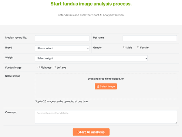
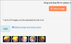
Up to 20 images can be uploaded as a batch
-
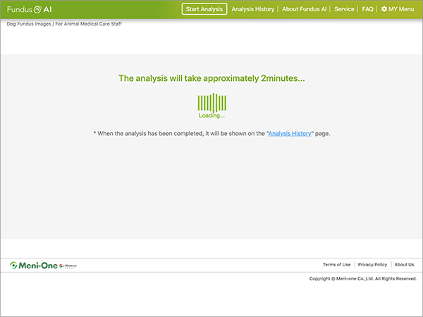
Analysis takes just a few minutes
-
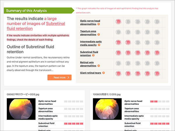
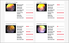
Possible similarities with each abnormal finding are displayed on graphs
-
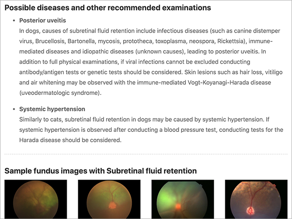
Detailed specialist explanations on each abnormal
finding are also displayed
-
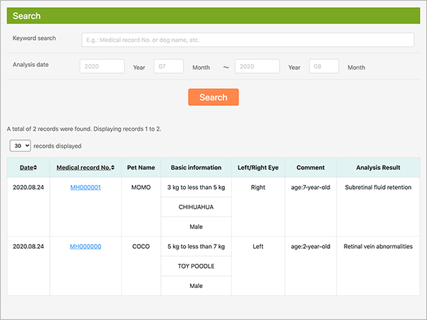
Analysis history is saved and can be checked later
-
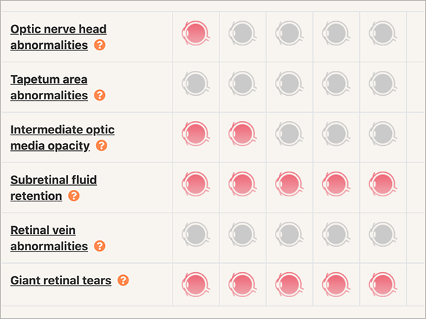
<Analyzed abnormal findings>
・Optic nerve head abnormalities・Tapetum area abnormalities
・Intermediate optic media opacity・Subretinal fluid retention
・Retinal vein abnormalities・Giant retinal tears
How to Use
Fundus images analyzed in three simple steps
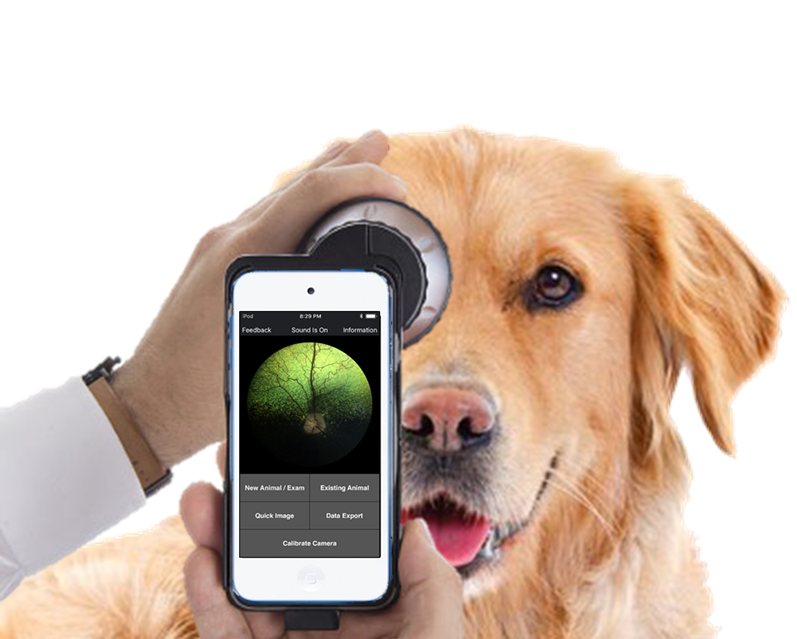
Photograph fundus with retinal camera
During the medical examination, the dog's fundus is photographed with a retinal camera. The fundus images actually photographed at the clinical practice can be used to run analyses related to ophthalmic findings.
Start analysis
Photographed fundus images are uploaded to Fundus AI. The analysis process will start.
Analysis complete
The analysis is completed on the spot. The degree of similarity with ophthalmic findings is displayed in the analysis results. Past analysis history can also be displayed.
Specialist explanations on each ophthalmic finding are also available for reference. This information is useful for subsequent examinations and determining treatment courses. It can also be printed out straight away and handed to the owner.

Specialist Feedback
Fully supervised by ophthalmologists

Seiya Maehara, Director, HIKARIMACHI Animal Eye Clinic
Naoaki Takiyama, Director, Veterinary Eye Clinic Nagoya

About Fundus AI
For inquiry
E-mail: support@ai-en.meni-one.com
Meni-one Co., Ltd.
Miyuki Business Park Building No. 4, 390 Ichibagi-cho, Nishi-ku, Nagoya, Aichi 452-0805 JAPAN